
Multiple Pressures Involved In Glaucoma
Cerebrospinal fluid ("CSF") fills the subarachnoid space in the brain and spinal cord, and bathes the optic nerve all the way to its insertion through the lamina cribrosa at the back of the eye. CSFp is the pressure of the cerebrospinal fluid (analogous to blood pressure or intraocular pressure or any fluid pressure).
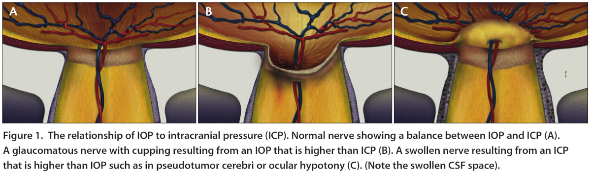
The intraocular pressure ("IOP") is separated from the CSFp by the lamina cribrosa, which is roughly 500 μm thick but thins with glaucoma. Any change in one of these pressures can be associated with a disturbance of homeostasis of the optic nerve head, such as glaucomatous optic neuropathy.
If the IOP is high or if the CSFp is low, then a pressure differential exists across the optic nerve head, and glaucomatous damage can occur. The relatively low CSFp or relatively elevated IOP will generate a net force on the optic nerve, causing posterior bowing of the lamina cribrosa and the cupping that is characteristic in eyes with glaucoma. (Figure 1).
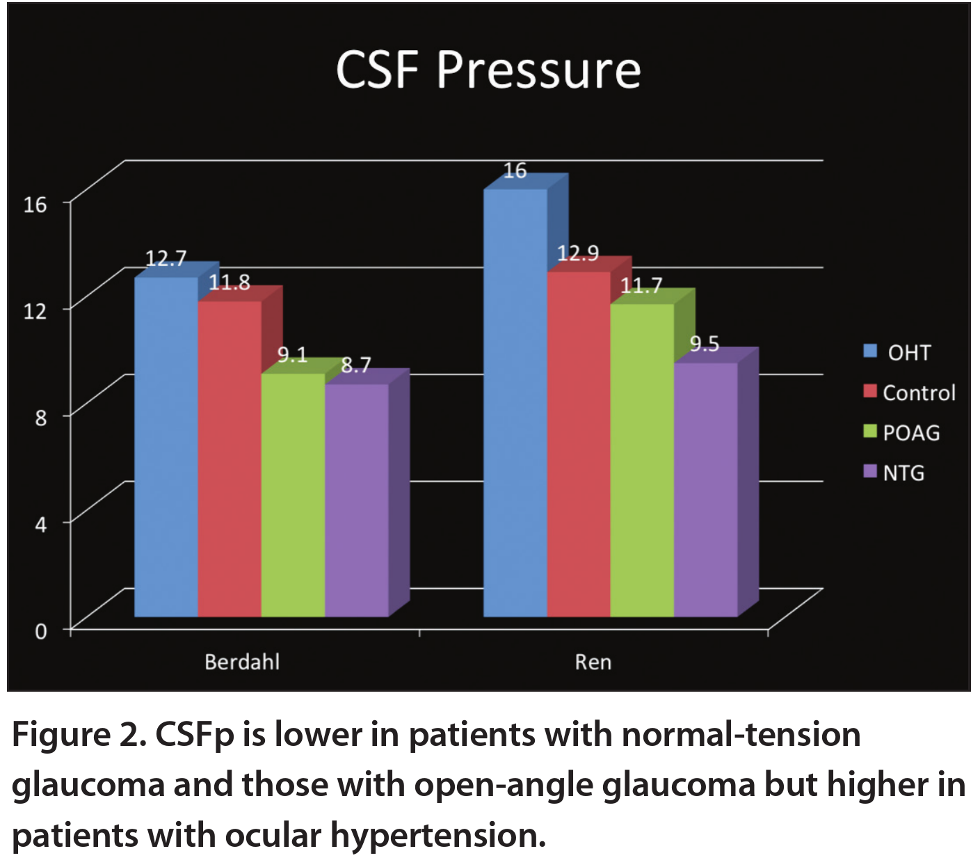
Studies have shown that patients with glaucoma have a lower CSFp, whereas those with ocular hypertension have a higher and possibly protective CSFp (Figure 2).
Based on the triangular relationships between IOP, CSFp, and blood pressure, glaucoma may be described as an imbalance between these three pressure parameters, eventually leading to an increased trans-lamina cribrosa pressure difference (TLCPD). 4
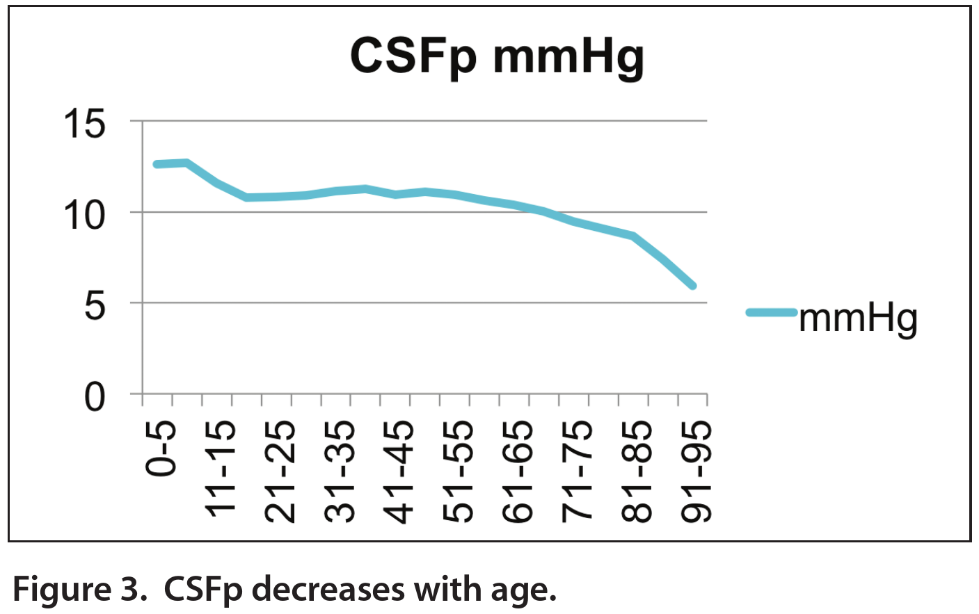
Other research has demonstrated a decrease in CSFp with increasing age, consistent with the rising incidence of glaucoma with increasing age (Figure 3). In the next section we will explore whether there are any lifestyle factors that glaucoma patients could employ to support a healthy balance between all these pressures.
Possible Complementary and Integrative Health Approaches
The meninges reside under the skull, where they protect the central nervous system and contain immune cells critical for the brain. The subarachnoid (SA) space in the meninges holds cerebrospinal fluid (CSF). The glymphatic (glial-lymphatic) system is an important clearance pathway for CSF, which flows into the brain via the para-arterial space, exchanges fluid contents with interstitial fluid (ISF), and then drains back to the SA space via the paravenous space.
The meningeal lymphatic system connects with the glymphatic system to transport the drained CSF/ISF from the SA space to the peripheral deep cervical lymph nodes for clearance.
Together, the two systems play a critical role in removing solutes and waste products, such as β-amyloid (Aβ), tau, lactate, and proinflammatory cytokines, from the brain. Functionally, the glymphatic system also contributes to brainwide nutrient delivery, whereas the meningeal lymphatic system shares similar functions as the peripheral lymphatic system in carrying out tissue homeostasis and immune trafficking/surveillance functions.
The glymphatic system has been implicated in several medical conditions. For instance, glymphatic clearance is suppressed in Alzheimer’s disease (AD), cortical spreading depression–associated migraine aura, traumatic brain injury (TBI), subarachnoid hemorrhage/ischemic stroke, multiple microinfarcts, diabetes-associated cognitive impairment, chronic stress, high-dose alcohol use, sleep deprivation, and the aging brain.
Similarly, experimental disruption of the meningeal lymphatic system exacerbates Aβ deposition and cognitive impairment in mice. Molecules such as vascular endothelial growth factor C (VEGF-C) have been shown to enhance cognitive function of aging mice by promoting meningeal lymphatic flow. Therefore, understanding how the glymphatic/lymphatic systems are regulated may help in the development of therapeutic strategies to prevent or delay a variety of diseases and disorders, including brain dysfunction and glaucoma.
The glymphatic and lymphatic systems have been shown to be highly regulated by many factors and several complementary approaches. For example, the glymphatic system is regulated by the sleep-awake state and is predominantly active in rodents during sleep. In addition, glymphatic flow can be partly driven by cerebral arterial pulsation, whereas lymph flow is primarily propelled by the surrounding smooth muscle cell contraction of the collecting lymphatic vessels. Multiple non-pharmacologic approaches have been shown to regulate brain lymphatic flows, such as:
- physical exercise
- continuous theta burst stimulation (cTBS),
- deep inhalation–mediated thoracic pressure reduction
- certain breathwork (pranayama) practices
Furthermore, osteopathic lymphatic pump manipulations and lymphatic drainage massage can regulate peripheral lymphatic flows.
Dietary supplements such as omega-3 polyunsaturated fatty acids have been shown to promote amyloid-β clearance through facilitation of aquaporin-4 (AQP4)-dependent glymphatic function in mice.
However, significant knowledge gaps remain in evaluating whether many other complementary approaches, including acupuncture, yoga, and meditation, may also modulate the lymphatic system in the central nervous system and/or peripheral tissues. More research is needed.
A thorough understanding of the mechanistic interactions between these approaches and the lymphatic and glymphatic systems will help us optimize therapeutic strategies and potentials for disease treatment, disease prevention, and promotion of health and well-being.